Sign In
Upload
Manuals
Brands
GE Manuals
Medical Equipment
Vivid S6
GE Vivid S6 Manuals
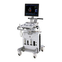
Manuals and User Guides for GE Vivid S6. We have
2
GE Vivid S6 manuals available for free PDF download: User Manual, Technical Publication
GE Vivid S6 User Manual (690 pages)
Brand:
GE
| Category:
Medical Equipment
| Size: 21.3 MB
Table of Contents
Table of Contents
3
Revision History
17
Attention
19
Prescription Device
19
Safety
19
Principles of Operation
20
Interference Caution
20
Indications for Use
21
Contraindications
21
Documentation
21
Manual Contents
22
Conventions Used in this Manual
23
Regulatory Requirements
24
Contact Information
25
Chapter 1 Safety
31
Introduction
33
Hazard Symbols
34
Owner Responsibility
35
Important Safety Considerations
36
Notice against User Modification
36
Regulatory Information
37
Directives
37
Product Classifications
37
Conformity to Standards
37
Certifications
39
Software License Acknowledgements
39
Device Labels
40
Label Locations
40
Label Icon Description
41
Acoustic Output
44
Definition of the Acoustic Output Parameters
44
Alara
44
Safety Statement
45
System Controls Affecting Acoustic Output
45
OB Exam
47
Exam Preparation
47
Acoustic Output Considerations
48
Concerns Surrounding Fetal Exposure
48
Patient Safety
49
Patient Identification
49
Diagnostic Information
49
Patient Guidance
50
Probe Safety
50
Mechanical Hazards
50
Electrical Hazard
51
Biological Hazards
52
Personnel and Equipment Safety
53
Explosion Hazard
53
Electrical Hazard
53
Smoke and Fire Hazard
54
Biological Hazard
54
Pacemaker Hazard
54
LCD Monitor
55
Electrical Safety
57
Internally Connected Peripheral Devices
57
External Connection of Other Peripheral Devices
57
Allergic Reactions to Latex-Containing Medical Devices
58
Use of ECG
59
Use of Defibrillator
59
Use of Electrosurgical Unit
59
Electromagnetic Compatibility (EMC)
60
EMC Performance
61
Declaration of Emissions
62
Declaration of Immunity
62
Notice Upon Installation of Product
62
General Notice
63
Peripheral Update for EC Countries
64
Patient Environmental Devices
66
Acceptable Devices
67
Unapproved Devices
67
Accessories, Options, and Supplies
67
Environmental Protection
68
Chapter 2 Getting Started
69
Introduction
70
Preparing the Unit for Use
71
Site Requirements
71
Connecting the Unit
72
Switching On/Off
79
Moving and Transporting the Unit
82
Wheels
82
Moving the Unit
82
Transporting the Unit
84
Reinstalling at a New Location
85
Preparing VIVID S5/VIVID S6 for Scanning
85
Unit Acclimation Time
86
System Description
87
System Overview
87
Control Panel
89
The Scanning Screen
102
Three-Pedal Footswitch Operation
105
Connecting and Disconnecting Probes
105
Adjusting the Display Monitor
108
Starting an Examination
113
Creating a New Patient Record or Starting an Examination from an Existing Patient Record
113
Selecting a Probe and an Application
118
Chapter 3 Basic Scanning Operations
121
Assignable Keys and Soft Menu Rocker
121
Using the Assignable Keys Soft Menu
122
Using the Soft Menu Rocker
125
Trackball Operation
126
Trackball Assignment
126
The System Menu
127
Cineloop Operation
128
Cineloop Overview
128
Cineloop Controls
129
Using Cineloop
130
Storing Images and Cineloops
131
To Store a Single Image
131
To Store a Cineloop
131
Removable Media
132
Intended Use
132
Supported Removable Media
133
Zoom
138
To Magnify an Image (Display Zoom)
138
To Activate the HR Zoom
138
Performing Measurements
139
To Perform Measurements
139
Physiological Trace
140
Connecting the Internal ECG
140
Physio Controls
144
Displaying the ECG Trace
144
Adjusting the Display of the ECG Trace
144
Annotations
146
To Insert an Annotation
146
To Edit Annotation
149
To Erase Annotation
149
Configuration of the Pre-Defined Annotation List
150
Bodymarks
152
Chapter 4 Scanning Modes
157
Introduction
157
2D-Mode
158
2D-Mode Overview
158
2D-Mode Controls
160
Using 2D
166
Optimizing 2D
166
M-Mode
168
M-Mode Overview
168
M-Mode Controls
169
Using M-Mode
171
Optimizing M-Mode
173
Color Mode
175
Color Mode Overview
175
Color M-Mode Overview
176
Color Mode Controls
177
Using Color Mode
180
Optimizing Color Mode
181
PW and CW Doppler
182
PW and CW Doppler Overview
182
PW and CW Doppler Controls
183
Using PW/CW Doppler Modes
185
Optimizing PW/CW Doppler Modes
186
Tissue Velocity Imaging (TVI)
187
Tissue Tracking
191
Strain Rate
196
Strain Rate Overview
196
Strain Rate Controls
197
Using Strain Rate
199
Optimizing Strain Rate
199
Strain
201
Strain Overview
201
Strain Controls
202
Using Strain
204
Optimizing Strain
204
Tissue Synchronization Imaging (TSI)
206
TSI Overview
206
TSI Controls
207
Using TSI
209
Optimizing TSI
210
Additional Scanning Features
211
Logiqview
211
Compound
212
B-Flow
213
Blood Flow Imaging
213
Virtual Convex
214
Chapter 5 Stress Echo
215
Introduction
216
Selection of a Stress Test Protocol Template
217
Image Acquisition
218
Starting Acquisition
219
Continuous Capture Mode
223
Analysis
231
Quantitative TVI Stress Echo Analysis
236
Accessing QTVI Stress Analysis Tools
237
Vpeak Measurement
238
Tissue Tracking
241
Quantitative Analysis
241
References
242
Editing/Creating a Template
243
Entering the Template Editor Screen
243
Template Editor Screen Overview
244
Editing/Creating a Template
247
Chapter 6 Contrast Imaging
251
Introduction
252
Cardiac Imaging
252
Non-Cardiac Imaging
253
Data Acquisition
253
Left Ventricular Contrast Imaging
253
LV Contrast Overview
254
LV Contrast Controls
254
Using LV Contrast
256
Optimizing LV Contrast
257
Vascular Contrast Imaging
258
Abdominal Contrast Imaging
259
Chapter 7 Measurement and Analysis
264
Introduction
264
About Measurement Results Display
265
The Assign and Measure Modality
266
Starting the Assign and Measure Modality
266
Entering a Study and Performing Measurements
267
Measure and Assign Modality
269
Starting the Measure and Assign Modality
269
Post-Measurement Assignment Labels
270
Cardiac Measurements
273
2D Measurements
273
M-Mode Measurements
277
Doppler Measurements
280
TSI Measurements
284
Automated Function Imaging
291
Autoef Measurements
310
Vascular Measurements
320
B-Mode Measurements
320
Intima-Media Thickness
321
M-Mode Measurements
326
Doppler Measurements
327
Pediatric Calculations
332
Overview
332
Hip Dysplasia Calculation
333
Making Hip Dysplasia Measurement
333
Performing an ob Exam
335
Patient Entry
335
Selecting Probe and ob Application
339
OB Measurements and Calculations
340
Introduction
340
B-Mode Measurements
341
M-Mode Measurements
348
Doppler Mode Measurements
348
OB Parameter Configuration
351
Configuring ob M&A According to Geographical Regions
351
Measurement Package Configuration
356
Measurement Package Configuration - Example
356
User-Defined Formulas
361
User-Defined Formula - Example
361
About Units
367
Measurement Result Table
369
Minimizing the Measurement Result Table
369
Moving the Measurement Result Table
369
Deleting Measurements
370
Worksheet
371
Overview
371
Using Worksheet
372
OB Worksheet
374
Patient Data
375
Measurement Information
375
Calculation Information
376
OB Graphs
377
Overview
377
Fetal Growth Curve Graph
378
Fetal Trending
382
Fetal Growth Bar Graph
383
OB-Multigestational
384
Multiple Fetus
384
GYN Measurements
388
Introduction
388
To Start a Gynecology Exam
388
B-Mode Measurements
389
Uterus Length, Width, and Height
389
Ovary Length, Width, and Height
390
Follicle Measurements Length, Width, and Height
391
Endometrium Thickness (Endo)
391
M-Mode Measurements
392
Doppler Mode Measurements
392
Chapter 8 Quantitative Analysis
395
Introduction
395
Accessing the Quantitative Analysis Package
396
In Replay Mode
396
In Live
396
Quantitative Analysis Window
397
Overview
397
Generation of a Trace
404
About the Sample Area
404
To Generate a Trace
404
Manual Tracking of the Sample Area (Dynamic Anchored Sample Area)
405
Zooming in the Analysis Window
406
Deletion of a Trace
407
To Delete All Traces
407
To Delete One Specific Trace
407
Saving/Retrieving Quantitative Analysis
407
Frame Disabling
408
Disabling Frames
408
Re-Enabling All Frames
408
Optimizing Sample Area
410
Reshaping a Sample Area
410
Labelling a Sample Area
411
Optimizing the Trace Display
412
Optimizing the Y-Axis
412
Trace Smoothing
413
Switching Modes or Traces
415
To Switch Mode
415
To Switch Trace
415
Cine Compound
416
Anatomical M-Mode
417
Introduction
417
Using Anatomical M-Mode
417
Optimizing Anatomical M-Mode
419
Chapter 9 Archiving
423
Introduction
423
Storing Images and Cineloops
424
Storing an Image
425
Storing a Cineloop
425
Saving Images and Cineloops to a Standard Format
426
Mpegvue/Evue
428
Retrieving and Editing Archived Information
431
Locating a Patient Record
431
Selecting a Patient Record and Editing Data in the Archive
436
Deleting Archived Information
440
Moving Examinations
442
Review Images in Archive
444
Review the Images from a Selected Examination
444
Select Images from the Image List Screen
445
Connectivity
449
The Dataflow Concept
449
Stand-Alone Scanner Scenario
452
A Stand-Alone Scanner and a Stand-Alone Echopac PC Environment
453
A Scanner and Echopac PC in a Direct Connect Environment
455
A Scanner and Echopac PC in a Network Environment
459
A Scanner and a DICOM Server in a Network
461
Export/Import Patient Records/Examinations
470
Exporting Patient Records/Examinations
470
Importing Patient Records/Examinations
478
Disk Management
482
Configuring the Disk Management Function
483
Running the Disk Management Function
486
Data Backup and Restore
489
DICOM Spooler
497
Starting the DICOM Spooler
497
Database Import from VIVID 3 or VIVID 4
500
Transfer Procedure
500
Installing the VIVID 3/4 Data Viewer
503
Using the VIVID 3/4 Data Viewer
506
Chapter 10 Report
510
Introduction
510
Creating a Report
511
Working with the Report Function
511
To Print a Report
514
To Store a Report
514
Retrieving an Archived Report
515
Deleting an Archived Report
515
Structured Findings
516
Prerequisite
516
Starting Structured Findings
517
Structured Findings Structure
517
Using Structured Findings
519
Structured Findings Configuration
522
Direct Report
532
Creating Comments
532
Creating Pre-Defined Text Inputs
533
Report Designer
534
Accessing the Report Designer
534
Report Designer Overview
534
Designing a Report Template
537
Saving the Report Template
548
To Exit the Report Designer
548
Report Templates Management
549
Configuration of the Template Selection Menu
550
Export/Import of Report Templates
551
Chapter 11 Probes
555
Probe Overview
555
Supported Probes
555
VIVID S5 Probe/Application Overview
560
VIVID S6 Probe/Application Overview
561
Maximum Probe Temperature
562
Probe Orientation
563
Probe Labelling
564
Environmental Requirements
565
Probe Integration
566
Selecting Probes
566
Connecting the Probe
566
Activating the Probe
568
Disconnecting the Probe
569
Care and Maintenance
570
Planned Maintenance
570
Inspecting the Probe
571
Special Handling Instructions
572
Cleaning and Disinfecting Probes
574
Probe Safety
580
Biopsy
581
Precaution Concerning the Use of Biopsy Procedures
581
Preparing the Biopsy Guide Attachment
583
Displaying the Guide Zone
587
Endocavitary Probe Biopsy Guide Assembly
589
Biopsy Needle Path Verification
591
Starting the Biopsy Procedure
591
Cleaning, Disinfection and Disposal
591
Surgery/Intra-Operative Use
592
Preparing for Surgery/Intra-Operative Procedures
592
Chapter 12 Peripherals
594
Introduction
594
Printing
595
To Print an Image
595
Specifications for Peripherals
595
DVR (Digital Video Recorder)
596
Overview
596
Using the DVR
596
Configuring the DVR
599
Reviewing the DVR Media Externally
599
Chapter 13 Presets and System Setup
601
Introduction
603
Starting the Configuration Package
606
To Open the Configuration Package
606
Overview
607
Imaging
608
The Global Setup Sheet
608
Application
611
Application Menu
614
Measure Text
616
The Measurement Menu Sheet
616
Configuration of the Measurement Menu
619
The Advanced Sheet
621
Parameter Configuration
621
The Modify Calculations Sheet
622
Parameter Configuration
622
The ob Table Sheet
623
Report
629
The Diagnostic Codes Sheet
630
The Comment Texts Sheet
631
Connectivity
634
Dataflow
634
Additional Outputs
641
Tools
643
Formats
644
Tcp-Ip
650
System
651
The System Settings
651
About
653
Administration
654
Users
655
Unlock Patient
658
Chapter 14 User Maintenance
660
System Care and Maintenance
660
Inspecting the System
660
Cleaning the Unit
661
Prevention of Static Electricity Interference
663
System Self-Test
664
System Malfunction
664
Using Insite Exc
668
Insite Exc Functionalities
668
Initiating a Request for Service (RFS)
668
Clinical Lifeline Mode
671
Exiting Insite Exc
672
Advertisement
GE Vivid S6 Technical Publication (127 pages)
Ultrasound System
Brand:
GE
| Category:
Medical Equipment
| Size: 3.71 MB
Table of Contents
Table of Contents
21
Chapter 1 - Overview
25
Content in this Manual
26
Product Description
27
Overview of the Ultrasound System
27
Products Covered in this Manual
28
Chapter 2 - Safety Information
29
Overview
30
Contents in this Chapter
30
Important Conventions
31
Conventions Used in Book
31
Standard Hazard Icons
33
Product Icons
35
Label Icon Description
35
How to Lock the Operator Panel Prior to Transport
35
Safety Considerations
36
Introduction
36
Human Safety
36
Mechanical Safety
39
Electrical Safety
44
Label Locations
46
Product Labels
46
Dangerous Procedure Warnings
47
Warnings
47
Lockout/Tagout (LOTO)
48
LOTO Requirements
48
Returning Probes and Repair Parts
49
Requirements for Returning Parts
49
User Responsibility
49
Electromagnetic Compatibility (EMC)
50
What Is EMC
50
Compliance
50
Electrostatic Discharge (ESD) Prevention
51
Chapter 3 - Site Preparations
53
Overview
54
Contents in this Chapter
54
General Ultrasound System Requirements
55
Contents in this Section
55
Ultrasound System Environmental Requirements
55
Electrical Requirements
57
EMI Limitations
70
Probes Environmental Requirements
72
Time and Manpower Requirements
72
Facility Needs
73
Contents in this Section
73
Purchaser Responsibilities
73
Required Facility Needs
75
Desirable Features
77
Care and Maintenance
79
Contents in this Chapter
80
Chapter 4 - Care and Maintenance
81
Warnings
81
Why Do Maintenance
82
Keeping Records
82
Quality Assurance
82
Maintenance Task Schedule
83
How Often Should Maintenance Tasks be Performed
83
System Maintenance
84
Preliminary Checks
84
Functional Checks
85
Physical Inspection
87
Cleaning
89
Probe Maintenance
90
Using a Phantom
91
Phantoms
91
Electrical Safety Tests
92
Content in this Section
92
Uninterrupted Power Supply (UPS)
93
Safety Test Overview
94
Leakage Current Limits
97
Outlet Test - Wiring Arrangement - USA and Canada
101
Grounding Continuity
102
Chassis Leakage Current Test
103
Isolated Patient Lead (Source) Leakage-Lead to Ground
107
Isolated Patient Lead (Source) Leakage-Lead to Lead
109
Isolated Patient Lead (Sink) Leakage-Isolation Test
111
Probe (Source) Leakage Current Test
113
Isolated Probe (Sink) Leakage-Isolation Test
118
When There's too Much Leakage Current
122
AC/DC Fails
122
Chassis Fails
122
Probe Fails
123
Peripheral Fails
123
Still Fails
123
New Ultrasound System
123
ECG Fails
123
Ultrasound Equipment Quality Check (EQC and IQC)
124
Quality Checks
124
Advertisement
Related Products
GE Vivid S5
GE VividS60
GE VividS70
GE Vivid 3 Pro
GE Vivid E9
GE VIVID 3 N RFI
GE Vivid in
GE Vivid FiVe
GE Vivid E9 Series
GE Vivid 7
GE Categories
Refrigerator
Ranges
Microwave Oven
Air Conditioner
Dishwasher
More GE Manuals
Login
Sign In
OR
Sign in with Facebook
Sign in with Google
Upload manual
Upload from disk
Upload from URL